What Is a Leg DVT Ultrasound?
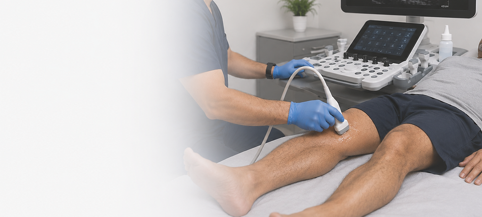
A Lower Extremity Venous DVT Ultrasound, also known as a Leg DVT Ultrasound or Venous Duplex Ultrasound, is a safe, noninvasive imaging examination used to evaluate the deep veins of one or both legs for the presence of a deep vein thrombosis (DVT), commonly known as a blood clot.
Using high-frequency sound waves, this examination allows your sonographer to assess blood flow and determine whether the veins are open and functioning normally. It is the preferred first-line imaging test for diagnosing or ruling out DVT because it is accurate, painless, and does not involve radiation.
A Lower Extremity Venous DVT Ultrasound is commonly performed when a patient experiences symptoms such as leg swelling, pain, tenderness, redness, warmth, or when there is concern for a blood clot based on medical history or risk factors.
Early diagnosis of DVT is important because an untreated blood clot can increase the risk of serious complications, including a pulmonary embolism, when part of the clot travels to the lungs.
Why Is a Lower Extremity Venous DVT Ultrasound Performed?
A Lower Extremity Venous DVT Ultrasound is performed to evaluate the deep and superficial veins of the legs for deep vein thrombosis (DVT) and other venous conditions. This examination provides detailed, real-time images of the veins while assessing blood flow without the use of radiation, contrast dye, or invasive procedures.
This examination is commonly performed when a blood clot is suspected due to symptoms such as leg swelling, pain, tenderness, redness, warmth, or one leg becoming noticeably larger than the other. It is also frequently used to evaluate patients with recent surgery, injury, prolonged immobility, pregnancy, cancer, or other conditions that increase the risk of developing a blood clot.
A Lower Extremity Venous DVT Ultrasound can identify blood clots, venous obstruction, and other abnormalities that may interfere with normal blood flow. It is also used to evaluate unexplained leg swelling, investigate abnormal blood test results such as an elevated D-dimer, and monitor patients with a history of deep vein thrombosis or other venous disorders.
Because ultrasound provides real-time imaging, it allows the technologist to assess both the structure of the veins and the direction of blood flow while confirming that the veins compress normally. This helps accurately diagnose or exclude the presence of a blood clot and guides timely treatment when necessary.
As a safe, painless, and radiation-free examination, a Lower Extremity Venous DVT Ultrasound is the preferred first-line imaging study for evaluating suspected blood clots and other venous conditions affecting the legs.
Common Reasons to Schedule a Lower Extremity Venous DVT Ultrasound
A Lower Extremity Venous DVT Ultrasound is performed to evaluate the veins of the legs for deep vein thrombosis (DVT) and other venous conditions. You may benefit from this examination if you are experiencing symptoms of a blood clot, have risk factors for DVT, or require follow-up of a known venous condition.
-
Swelling of one or both legs
-
Leg pain or tenderness
-
Redness or warmth of the leg
-
A feeling of heaviness or tightness in the leg
-
One leg that is noticeably larger than the other
-
Suspected deep vein thrombosis (DVT)
-
Evaluation of unexplained leg pain or swelling
-
Recent surgery, injury, or prolonged immobility
-
Pregnancy or the postpartum period with symptoms concerning for DVT
-
Elevated D-dimer blood test with concern for DVT
-
Personal or family history of blood clots
-
Medical conditions that increase the risk of blood clot formation
-
Cancer or ongoing cancer treatment
-
Follow-up evaluation of a previously diagnosed lower extremity DVT
-
Monitoring response to blood thinner (anticoagulant) therapy
-
Evaluation of abnormal findings from a physical examination or another imaging study
A Lower Extremity Venous DVT Ultrasound provides detailed, real-time images of the veins and evaluates blood flow to detect blood clots (deep vein thrombosis), venous obstruction, and other abnormalities. It is a safe, painless, and radiation-free examination that helps guide prompt diagnosis, treatment, and follow-up care.
What to Expect During Your Examination
A Lower Extremity Venous DVT Ultrasound is a safe, painless, and noninvasive examination that typically takes 30 to 45 minutes, depending on whether one or both legs are being evaluated.
During the examination, you will be asked to lie comfortably on an examination table while a clear, water-based ultrasound gel is applied to your skin. The gel helps transmit sound waves and allows clear images of your veins to be obtained.
Your sonographer will gently move an ultrasound transducer over your leg while evaluating the deep veins. Gentle pressure, known as compression, will be applied at several locations along the veins. This is a normal and essential part of the examination and is one of the most reliable ways to determine whether a blood clot is present.
Color Doppler and spectral Doppler ultrasound may also be used to evaluate blood flow throughout the veins. During the examination, you may be asked to slightly change your position or perform simple breathing maneuvers to help assess venous blood flow.
This examination does not use radiation, injections, or contrast dye. Most patients experience little to no discomfort, although mild tenderness may be felt if the affected area is already painful.
Once the examination is complete, you may immediately return to your normal daily activities unless your healthcare provider gives you different instructions.
How to Prepare for Your Lower Extremity Venous DVT Ultrasound
Preparing for your Lower Extremity Venous DVT Ultrasound is simple, and no special preparation is typically required. You may eat and drink normally and continue taking your regular medications unless you have been given different instructions.
If you have a history of deep vein thrombosis (DVT), recent leg surgery, leg trauma, venous procedures, or are currently taking blood-thinning medications, please let your sonographer know before your examination. It is also helpful to mention any current symptoms, such as leg swelling, pain, tenderness, redness, or warmth, as this information may assist with the evaluation.
Before Your Appointment
✓ Wear comfortable, loose-fitting clothing or shorts that allow easy access to your leg(s). You may be asked to change into a medical gown for the examination.
✓ Continue taking your prescribed medications unless you have been instructed otherwise.
✓ You may eat and drink normally before your appointment unless you have received different instructions.
✓ Avoid applying lotions, oils, or creams to your legs on the day of your examination, as they may interfere with the ultrasound gel and image quality.
✓ Bring any relevant medical records or previous ultrasound reports if they are available and have not already been provided.
✓ Arrive 10–15 minutes early to complete any necessary paperwork and check in before your scheduled appointment.
A Lower Extremity Venous DVT Ultrasound requires no injections, contrast dye, radiation, or special preparation, making it a simple, safe, and convenient examination. Following these instructions will help ensure your examination is performed as comfortably and efficiently as possible.
Frequently Asked Questions
Q: What is a deep vein thrombosis (DVT)?
A: A deep vein thrombosis (DVT) is a blood clot that forms in one of the deep veins, most commonly in the leg. If left untreated, part of the clot can break free and travel to the lungs, causing a pulmonary embolism, a potentially life-threatening condition.
Q: Is a Lower Extremity Venous DVT Ultrasound painful?
A: No. The examination is noninvasive and generally painless. You may feel mild pressure as the sonographer compresses the veins with the ultrasound transducer. If your leg is already tender, you may experience slight discomfort during those portions of the examination.
Q: How long does the examination take?
A: Most examinations take approximately 30 to 45 minutes, depending on whether one or both legs are being evaluated and the complexity of the study.
Q: Do I need to prepare for the examination?
A: No special preparation is required. You may eat, drink, and take your usual medications unless you have been instructed otherwise.
Q: Will I be exposed to radiation?
A: No. Ultrasound uses high-frequency sound waves to create images and does not involve radiation.
Q: What should I wear for the examination?
A: Loose-fitting clothing or shorts are recommended so the leg being examined is easily accessible. A gown can be provided if needed.
Q: Can I drive and return to work afterward?
A: Yes. Most patients can return to their normal daily activities immediately after the examination.
Q: Can this examination detect all causes of leg pain or swelling?
A: This examination is specifically designed to evaluate the deep veins for blood clots. Although other findings, such as fluid collections or Baker's cysts, may occasionally be seen, additional imaging may be recommended if another condition is suspected.
Q: What happens if a blood clot is found?
A: If a significant blood clot is identified, the findings will be communicated promptly so that appropriate medical care can be arranged. Depending on the results, you may be advised to seek immediate evaluation or follow up with your healthcare provider.
Q: Can I schedule this examination without a referral?
A: Yes. Depending on your circumstances and applicable regulations, this examination may be scheduled as a self-pay service or following a referral from a healthcare provider. If you are experiencing severe symptoms such as sudden shortness of breath, chest pain, or significant leg swelling, seek immediate medical attention rather than delaying care for an outpatient examination.
Schedule Your Lower Extremity Venous DVT Ultrasound
Scheduling your Lower Extremity Venous DVT Ultrasound is quick and convenient. Whether your examination has been recommended by a healthcare provider or you are scheduling a self-pay evaluation, you can book your appointment online in just a few minutes.
Choose the option below that best fits your needs. If you need an ultrasound of one leg, select the One Leg examination. If you need both legs evaluated, select the Two Legs examination and proceed to schedule your appointment.
Our experienced registered vascular sonographer performs every examination using advanced ultrasound technology with a focus on diagnostic accuracy, patient comfort, and personalized care.
If you are experiencing sudden chest pain, shortness of breath, severe leg swelling, or other symptoms of a medical emergency, call 911 or seek immediate emergency medical care rather than scheduling an outpatient ultrasound examination.
Why Choose Alpha Vascular?
Experienced Sonographer

Board-Certified Interpretation

Advanced Ultrasound Technology
