What Is a Lower Extremity Venous Insufficiency Ultrasound?
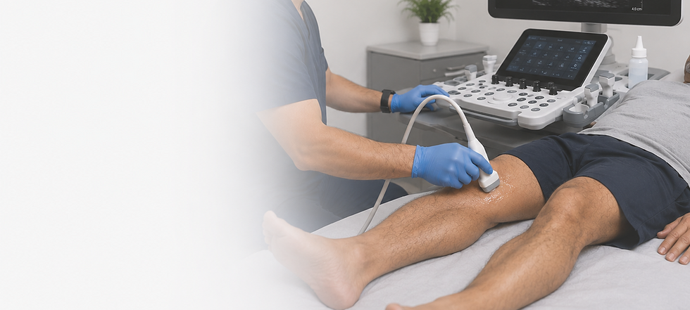
A Lower Extremity Venous Insufficiency Ultrasound, also known as a Venous Reflux Ultrasound or Venous Duplex Ultrasound, is a safe, noninvasive imaging examination used to evaluate the veins in one or both legs for venous insufficiency, a condition in which the vein valves do not function properly. When these valves become weakened or damaged, blood can flow backward (reflux) and pool in the veins instead of returning efficiently to the heart.
Using high-frequency sound waves, this examination allows your sonographer to evaluate both the anatomy of the veins and the direction of blood flow. During the study, specialized Doppler ultrasound techniques are used to identify venous reflux, assess valve function, and determine the severity and location of abnormal blood flow.
A Lower Extremity Venous Insufficiency Ultrasound is commonly performed when an individual experiences symptoms such as leg heaviness, aching, swelling, fatigue, varicose veins, skin discoloration, or nonhealing venous ulcers. It is also used to evaluate patients before vein treatments and to help determine the most appropriate treatment plan.
Because this examination identifies the underlying cause of chronic venous disease, it plays an important role in diagnosing venous insufficiency, guiding treatment decisions, and monitoring the effectiveness of vein procedures over time.
Why Is a Lower Extremity Venous Insufficiency Ultrasound Performed?
A Lower Extremity Venous Insufficiency Ultrasound is performed to evaluate the veins of the legs and determine whether the valves within the veins are functioning properly. This examination provides detailed, real-time images of the veins while assessing blood flow and identifying venous reflux (backward flow of blood) without the use of radiation, contrast dye, or invasive procedures.
This examination is commonly performed to investigate symptoms of chronic venous insufficiency, including leg pain, aching, heaviness, fatigue, swelling, varicose veins, skin discoloration, or venous ulcers. It helps determine whether these symptoms are caused by malfunctioning vein valves that allow blood to flow backward and pool in the legs.
A Lower Extremity Venous Insufficiency Ultrasound evaluates both the superficial and deep venous systems, measuring the direction and duration of blood flow to identify areas of venous reflux. It can also detect enlarged varicose veins, incompetent perforator veins, and other abnormalities that may contribute to chronic venous disease.
Because ultrasound provides real-time imaging, the examination is often performed with you standing or in a reverse Trendelenburg position to accurately evaluate vein valve function under normal physiologic conditions. The technologist may also use gentle compression and specialized maneuvers to assess blood flow and valve competency.
As a safe, painless, and radiation-free examination, a Lower Extremity Venous Insufficiency Ultrasound is the preferred first-line imaging study for evaluating chronic venous disease, guiding treatment decisions, and determining whether procedures such as endovenous ablation, sclerotherapy, or other vein treatments may be beneficial.
Common Reasons to Schedule a Lower Extremity Venous Insufficiency Ultrasound
A Lower Extremity Venous Insufficiency Ultrasound is performed to evaluate the veins of the legs for venous reflux (backward blood flow) and other conditions affecting the venous circulation. You may benefit from this examination if you are experiencing symptoms of chronic venous insufficiency, have visible varicose veins, or require evaluation before or after vein treatment.
-
Varicose veins
-
Leg pain or aching
-
A feeling of heaviness or fatigue in the legs
-
Leg swelling
-
Burning, throbbing, or cramping in the legs
-
Leg discomfort that worsens after prolonged standing or sitting
-
Visible enlarged or twisted veins
-
Skin discoloration around the ankles or lower legs
-
Venous ulcers or slow-healing wounds
-
Itching or irritation over varicose veins
-
Restless or tired legs associated with venous disease
-
Suspected chronic venous insufficiency
-
Evaluation before endovenous ablation, sclerotherapy, or other vein treatments
-
Follow-up after vein treatment or surgery
-
Evaluation of recurrent varicose veins
-
Evaluation of abnormal findings from a physical examination or another imaging study
A Lower Extremity Venous Insufficiency Ultrasound provides detailed, real-time images of the veins while evaluating vein valve function and detecting venous reflux. It is a safe, painless, and radiation-free examination that helps diagnose chronic venous disease, guide treatment decisions, and monitor treatment outcomes.
What to Expect During Your Examination
A Lower Extremity Venous Insufficiency Ultrasound is a safe, painless, and noninvasive examination that typically takes 45 to 60 minutes, depending on whether one or both legs are being evaluated.
Unlike a DVT ultrasound, this examination is specifically designed to evaluate venous valve function and blood flow. To obtain the most accurate results, much of the examination is performed while you are standing or positioned on a tilt table, allowing gravity to demonstrate whether blood is flowing backward (venous reflux).
A clear, water-based ultrasound gel will be applied to your skin, and your sonographer will move an ultrasound transducer along the veins of your leg while obtaining detailed images. Gentle compression and a series of breathing and compression maneuvers may be performed to evaluate the direction of blood flow and measure the duration of venous reflux.
Throughout the examination, multiple superficial and deep veins will be evaluated to determine which veins are affected and the severity of any venous insufficiency. This information is essential for developing the most appropriate treatment plan.
The examination does not use radiation, injections, or contrast dye. Most patients experience little to no discomfort and may return to their normal daily activities immediately after the examination.
How to Prepare for Your Lower Extremity Venous Insufficiency Ultrasound
Preparing for your Lower Extremity Venous Insufficiency Ultrasound is simple, and no special preparation is typically required. You may eat and drink normally and continue taking your regular medications unless you have been given different instructions.
If you have had previous vein treatments, such as endovenous ablation, sclerotherapy, vein stripping, phlebectomy, or other venous procedures, please let your sonographer know before your examination. It is also helpful to mention any history of deep vein thrombosis (DVT), leg surgery, leg injury, or current symptoms such as leg heaviness, swelling, aching, varicose veins, or skin changes.
Before Your Appointment
✓ Wear comfortable clothing, such as shorts or loose-fitting pants, that allows easy access to your leg(s). You may be asked to change into a medical gown for the examination.
✓ Continue taking your prescribed medications unless you have been instructed otherwise.
✓ You may eat and drink normally before your appointment unless you have received different instructions.
✓ Avoid applying lotions, oils, or creams to your legs on the day of your examination, as they may interfere with the ultrasound gel and image quality.
✓ Because portions of the examination are performed while standing, wear comfortable footwear and let your sonographer know if you have difficulty standing for extended periods.
✓ Bring any relevant medical records or previous vein ultrasound reports if they are available and have not already been provided.
✓ Arrive 10–15 minutes early to complete any necessary paperwork and check in before your scheduled appointment.
A Lower Extremity Venous Insufficiency Ultrasound requires no injections, contrast dye, radiation, or special preparation, making it a simple, safe, and convenient examination. Following these instructions will help ensure your examination is performed as comfortably and efficiently as possible.
Frequently Asked Questions
Q: What is a Lower Extremity Venous Insufficiency Ultrasound?
A: A Lower Extremity Venous Insufficiency Ultrasound is a noninvasive examination that evaluates the veins in one or both legs for venous reflux, a condition in which the vein valves do not function properly. The examination helps determine whether blood is flowing backward and pooling in the veins.
Q: Is a Lower Extremity Venous Insufficiency Ultrasound painful?
A: No. The examination is generally painless. You may feel mild pressure from the ultrasound transducer or during compression maneuvers, but most patients experience little to no discomfort.
Q: How long does the examination take?
A: Most examinations take approximately 45 to 60 minutes, depending on whether one or both legs are being evaluated and the complexity of the findings.
Q: Will I need to stand during the examination?
A: Yes. Because gravity helps reveal abnormal blood flow, much of the examination is performed while you are standing or positioned on a tilt table. This allows the sonographer to accurately evaluate venous valve function and detect reflux.
Q: Do I need to prepare for the examination?
A: No special preparation is required. You may eat, drink, and take your usual medications unless you have been instructed otherwise.
Q: Will I be exposed to radiation?
A: No. Ultrasound uses high-frequency sound waves to create images and does not involve radiation.
Q: Can this examination diagnose blood clots (DVT)?
A: This examination is specifically designed to evaluate venous insufficiency (venous reflux) rather than to diagnose an acute deep vein thrombosis (DVT). If a blood clot is suspected, a dedicated Lower Extremity Venous DVT Ultrasound is typically recommended.
Q: Can I return to my normal activities after the examination?
A: Yes. You may return to your normal daily activities immediately after the examination.
Q: What happens if venous insufficiency is found?
A: Your results will identify which veins are affected, the severity of the reflux, and whether treatment may be beneficial. These findings help determine the most appropriate management plan, which may include conservative measures, compression therapy, or vein treatment procedures.
Q: Can I schedule this examination without a referral?
A: Yes. Depending on your circumstances and applicable regulations, this examination may be scheduled as a self-pay service or following a referral from a healthcare provider.
Schedule Your Lower Extremity Venous Insufficiency Ultrasound
Scheduling your Lower Extremity Venous Insufficiency Ultrasound is quick and convenient. Whether your examination has been recommended by a healthcare provider or you are scheduling a self-pay evaluation, you can book your appointment online in just a few minutes.
Choose the option below that best fits your needs. If you need an ultrasound of one leg, select the One Leg examination. If both legs need to be evaluated, select the Two Legs examination and proceed to schedule your appointment.
Every examination is performed by an experienced registered vascular sonographer using advanced ultrasound technology to accurately evaluate venous valve function, identify venous reflux, and provide detailed information to help guide the most appropriate treatment plan.
If you have severe leg pain, sudden swelling, redness, warmth, or other symptoms concerning for an acute blood clot (DVT), seek immediate medical evaluation rather than scheduling a venous insufficiency ultrasound, as a dedicated Lower Extremity Venous DVT Ultrasound may be the more appropriate examination.
Why Choose Alpha Vascular?
Experienced Sonographer

Board-Certified Interpretation

Advanced Ultrasound Technology
